
Desmoplastic ameloblastoma of Maxilla

Ameloblastoma, a relatively common epithelial odontogenic tumor includes several histopathologic subtypes like follicular, plexiform, acanthomatous and desmoplastic variants. Hybrid desmoplastic ameloblastoma (DA) composed of typical desmoplastic ameloblastoma along with areas of follicular/plexiform ameloblastoma is an extremely rare variant of ameloblastoma.

       Desmoplastic ameloblastoma (DA) was first
described in detail by Eversole et al in 1984 and
is defined as “a variant of ameloblastoma with
specific clinical, imaging and histological                   
features” in the recent WHO classification of
odontogenic tumors. Thus, it often occurs in the
anterior region of jaws, presents with unique
radiographic appearance resembling fibrosseous
lesions and show distinct histopathology
characterized by extensive stromal
collagenisation or desmoplasia surrounding
compressed islands of odontogenic epithelium
making it a distinct entity.


Ameloblastoma is a rare odontogenic tumor accounting for around 1% of all the cysts and tumors in the jaws.It encompasses several histological variants like follicular, plexiform,basaloid,acanthomatous and desmoplastic variants.The striking difference in the anatomic location i.e. occurrence in the anterior-premolar region ofmaxilla/mandible, unusual radiologic presentation of mixed radiolucency-radiopacities with illdefined borders and distinctive histopathology of extensive stromal desmoplasia with scattered odontogenic epithelium makes it a distinct clinicopathologic entity. Additional findings
reported for DA are almost equal sex predilection and relative higher frequency of occurrence in
Asians.


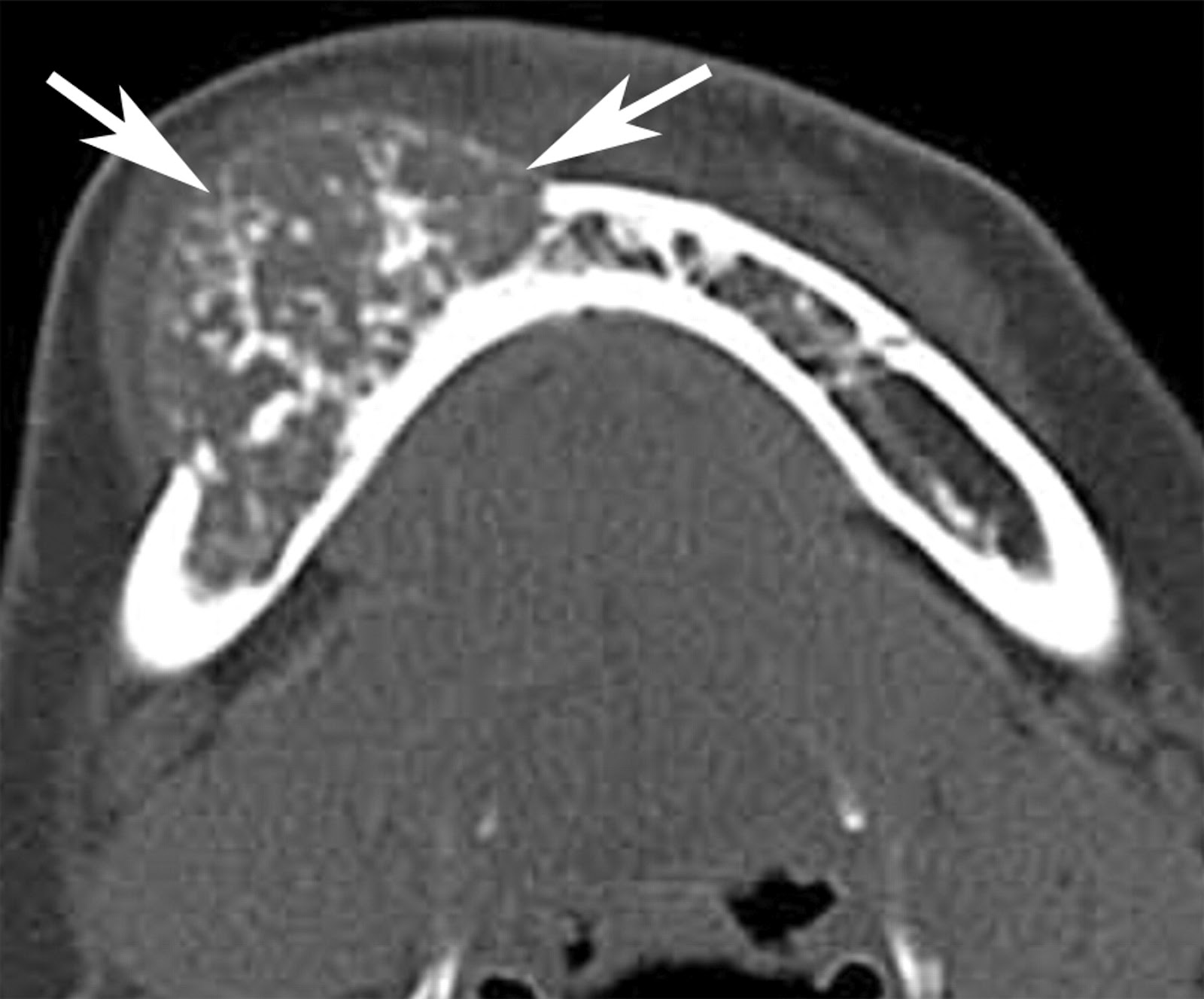
A: Intraoral clinical photograph of the tumor in the
left maxilla. B, PNS view demonstrating a mixed
radiolucent and radiopaque appearance with opacification of
the sinus. Computed tomography scan demonstrating a mixed
radiolucent –radiopaque lesion (C). Cut surface of the gross
specimen demonstrating a solid, granular, creamish white
lesion (D). E: Radiograph of the gross specimen depicting a
mottled appearance with focal radiolucencies.

For further reference :
Source : http://www.easternjmed.org/PDF/2011_1/9.pdf

Comments

Popular posts from this blog

How to use digital X-ray Or RVG with Apple iPad , iPhone. The Kodak RVG 6500

iPad Innovation is the key to development and dentistry is not an exception.First there was no radiograph then came conventional radiograph then came high speed radiograph and Digital radiograph or RVG  and now Apple iPad. You will say ( What ! an iPad?)  yes an iPad. It's an innovation by Apple inc.which has been accepted by medical and dental field warmly.I have discussed How to upgrade your dental practice with Apple iPad in earlier post and written about the change this gadget can bring.You can check  10 free iPad application for dentists Today in Digital Radiograph or RVG we shoot an X-ray few moments later it appears on the computer screen and then you interpret it and discuss it with your patients. Now Kodak have made a RVG system which is iPad compatible and if you have an iPad or iPhone you can see and review this X-ray image directly on your gadget write reports and save it. You must have Two thing for it. 1.RVG Mobile software in your iPad...

The Calla lilly prep in endodontics !!

During patient treatment, the clinician needs to consider many factors that will affect the ultimate outcome. In simple terms, these factors can be grouped into 3 categories: (I) operator needs, (II) restoration needs, (III) the tooth needs. (I)The operator needs are the conditions the clinician needs to treat the tooth. (II) The restoration needs are the prep dimensions and tooth conditions for optimal strength and longevity. (III)The tooth needs are the biologic and structural limitations for a treated tooth to remain predictably functional. The Cala Lilly is a flower and is the new model for composite preparations. SOURCE: Modern Molar Endodontic Access and Directed Dentin Conservation, David Clark et al,2010

Articulator

Articulators    DEFINATION:  A Mechanical device that represents the temperomandibular joints and jaw members to which maxillary and mandibular casts may be attached to stimulate some or all of the mandibular movements. PURPOSES 1.   To  hold the maxillary and mandibular casts  in a determined fixed relationship. 2. Mounting of dental casts  for diagnosis treatment planning and patient presentation.   3.To simulate the jaw movement like  opening and closing. 4. Fabrication of  occlusal surfaces  for dental restoration. 5.  Arrangement of artificial teeth  for complete and removable partial denture.       USE      1. To diagnose the  state of occlusion in both  the natural and artificial dentition      2.   To plan the  dental procedures  based on the relationship between opposing natural and        ...