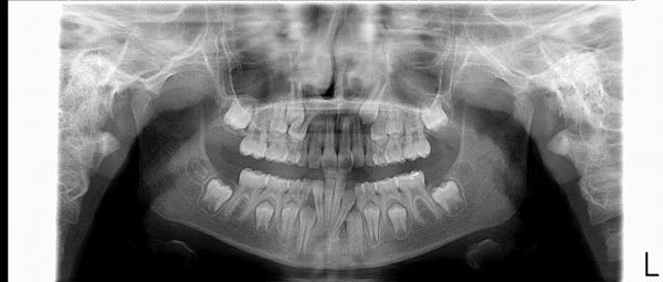
Here is a really good direct digital panoramic radiograph (courtesy of Planmeca). If you know what to look for, you can tell that this patient (probably 9 or 10 years old) is going to need orthodontic treatment!

In the last several years, systems have become available for digital radiographs. What does that mean? If it’s digital it must be better, right? Well, here are some advantages and differences:
1. The same x-ray machine is used to produce the image. It is possible to reduce the dosage administered with digital.
2. The image is no longer on a film, therefore no more developing chemicals or processing errors.
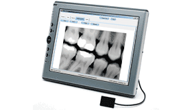
3. The image can be enlarged and enhanced for better viewing. I don’t know how many times I have showed a patient (or parent) decay between the teeth (obvious to me) on that little 1 inch square film and the patient says, yes I see that. Well, sometimes I wonder if they are just being nice, because it took me a long time to be able to read some of those films. I look at a thousand each day! Now I can make that little image as big as the computer screen.
4. You can more easily store, retrieve, compare, manipulate, enhance and e-mail radiographs.
5. Film still has the best quality of image in my opinion. It is hard to match the resolution of film, but things are improving.
6. There are direct CCD and phosphor plate systems. The phosphor plates are like film and retain an image when struck by the X-ray. The plate is then scanned for the data. Direct digital systems have a CCD sensor that takes the same X-ray and converts it directly into the data used for presentation and storage--no scanning needed but the sensor can be hard to place in the mouth of a child.
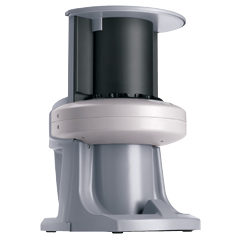
7. Digital Panoramic/Ceph systems are expensive! (more than $80,000 right now).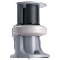
I have to say these digital systems are quirky. Haven't you had a problem with your computer? Well, the same kinds of things can happen with these systems. In addition to the scanner or digital CCD sensor, there is the software you use to manipulate and display the images and the office practice software, etc, etc.. Well, two steps forward, one back!
*(By the way, the reason there is an "L" on the X-ray is to orient the viewer as to what is the patient's left side. I often say it's like the patient is sitting in a chair smiling at us).
In the last several years, systems have become available for digital radiographs. What does that mean? If it’s digital it must be better, right? Well, here are some advantages and differences:
1. The same x-ray machine is used to produce the image. It is possible to reduce the dosage administered with digital.
2. The image is no longer on a film, therefore no more developing chemicals or processing errors.
3. The image can be enlarged and enhanced for better viewing. I don’t know how many times I have showed a patient (or parent) decay between the teeth (obvious to me) on that little 1 inch square film and the patient says, yes I see that. Well, sometimes I wonder if they are just being nice, because it took me a long time to be able to read some of those films. I look at a thousand each day! Now I can make that little image as big as the computer screen.

4. You can more easily store, retrieve, compare, manipulate, enhance and e-mail radiographs.
5. Film still has the best quality of image in my opinion. It is hard to match the resolution of film, but things are improving.
6. There are direct CCD and phosphor plate systems. The phosphor plates are like film and retain an image when struck by the X-ray. The plate is then scanned for the data. Direct digital systems have a CCD sensor that takes the same X-ray and converts it directly into the data used for presentation and storage--no scanning needed but the sensor can be hard to place in the mouth of a child.
7. Digital Panoramic/Ceph systems are expensive! (more than $80,000 right now).
I have to say these digital systems are quirky. Haven't you had a problem with your computer? Well, the same kinds of things can happen with these systems. In addition to the scanner or digital CCD sensor, there is the software you use to manipulate and display the images and the office practice software, etc, etc.. Well, two steps forward, one back!
*(By the way, the reason there is an "L" on the X-ray is to orient the viewer as to what is the patient's left side. I often say it's like the patient is sitting in a chair smiling at us).
Such a informative and full of knowledge article. Really appreciate your work. Hope to see these kind of articles in future.
ReplyDelete24 hour dentist omaha