A club foot, or congenital talipes equinovarus (CTEV), is a congenital deformity involving one foot or both. The affected foot appears rotated internally at the ankle. TEV is classified into 2 groups: Postural TEV or Structural TEV.
 .
.
Etiology
The true etiology of congenital clubfoot is unknown. Most infants who have clubfoot have no identifiable genetic, syndromal, or extrinsic cause.
Extrinsic associations include teratogenic agents (eg, sodium aminopterin), oligohydramnios, and congenital constriction rings. Genetic associations include mendelian inheritance (eg, diastrophic dwarfism; autosomal recessive pattern of clubfoot inheritance).
Cytogenetic abnormalities (eg, congenital talipes equinovarus [CTEV]) can be seen in syndromes involving chromosomal deletion. It has been proposed that idiopathic CTEV in otherwise healthy infants is the result of a multifactorial system of inheritance. Evidence for this is as follows:
- Incidence in the general population is 1 per 1000 live births.
- Incidence in first-degree relations is approximately 2%.
- Incidence in second-degree relations is approximately 0.6%.
- If one monozygotic twin has a CTEV, the second twin has only a 32% chance of having a CTEV.
Presentation
Seek a detailed family history of clubfoot or neuromuscular disorders, and perform a general examination to identify any other abnormalities. Examine the feet with the child prone, with the plantar aspect of the feet visualized, and supine to evaluate internal rotation and varus. If the child can stand, determine if the foot is plantigrade, if the heel is bearing weight, and if it is in varus, valgus, or neutral.
Similar deformities are seen with myelomeningocele and arthrogryposis. Therefore, always examine for these associated conditions. The ankle is in equinus, and the foot is supinated (varus) and adducted (a normal infant foot usually can be dorsiflexed and everted, so that the foot touches the anterior tibia). Dorsiflexion beyond 90° is not possible.
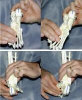
The navicular is displaced medially, as is the cuboid. Contractures of the medial plantar soft tissues are present. Not only is the calcaneus in a position of equinus but also the anterior aspect is rotated medially and the posterior aspect laterally.
The heel is small and empty. The heel feels soft to the touch (akin to the feel of the cheeks). As the treatment progresses, it fills in and develops a firmer feel (akin to the feel of the nose or of the chin).
The talar neck is easily palpable in the sinus tarsi as it is uncovered laterally. Normally, this is covered by the navicular, and the talar body is in the mortise. The medial malleolus is difficult to palpate and is often in contact with the navicular. The normal navicular-malleolar interval is diminished.
The hindfoot, as shown below, is supinated, but the foot is often in a position of pronation relative to the hindfoot. The first ray often drops to create a position of cavus. The Ponseti method of closed management of clubfeet through manipulations and casting describes the elevation of the first metatarsal as a first step, even if it means seemingly exacerbating the supination of the foot.
 Spontaneous correction of the hind foot varus by abducting the forefoot and allowing the calcaneum to freely rotate under the talus. The tibia often has internal torsion. This assumes special importance in the casting management of clubfeet, as shown in the images below, where care should be taken to rotate the feet into abduction, avoiding spurious tibial rotation through the knee.
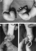 Never forcibly evert or pronate the foot during clubfoot casting.
 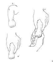 Traditional manipulation and casting methods fail, as they do not allow the free rotation of the calcaneum and the talus. Even following correction, the foot often remains short and the calf thin.
Indications
Traditionally, surgery is indicated when a plateau has been reached in nonoperative treatment. Surgery is usually performed when the child is of sufficient size to enable anatomy to be recognized.
Contraindications
No specific contraindications to surgery exist, although the child's size dictates that surgery is best performed at approximately age 6 months. With greater acceptance of the Ponseti conservative technique, surgery is seen to be a contentious issue. Surgery for clubfeet is no longer the only standard of care.
Comments
Post a Comment